Mr Joseph Aquilina, Consultant Obstetrician and Gynaecologist
Mr Joseph Aquilina
Consultant Obstetrician and Gynaecologist
Mr Joseph Aquilina FRCOG MFFP
Consultant Obstetrician and Gynaecologist
Mr Joseph Aquilina
Consultant Obstetrician and Gynaecologist FRCOG MFFP
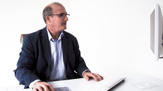
Mr Aquilina is a Consultant Obstetrician and Gynaecologist


Areas of expertise
- Endometriosis
- Fibroids
- PCOS
- Menopause
- Hysteroscopy and laparoscopic surgery

Make an appointment
Address
-
London Bridge Hospital
27 Tooley Street, London, SE1 2PR


-
HCA UK at The Shard
The Shard, 32 St Thomas Street, London, SE1 9BS
-
The Portland Hospital
205-209 Great Portland Street, London, W1W 5AH
About Mr Joseph Aquilina
GMC number: 4168337
Year qualified: 1987
Place of primary qualification: University of Malta
Initial consultation fee: £275
Follow up consultation fee: £195
Mr Joseph Aquilina is a Consultant Obstetrician and Gynaecologist with a wealth of experience in general gynaecology.
He specialises in treating women with menstrual disorders, polycystic ovarian syndrome, and pelvic pain. Additionally, he is highly skilled in foetal and maternal ultrasound. Mr Aquilina obtained his primary medical degree in Malta in 1987 and moved to the UK in 1989 for his postgraduate training.
He trained in Bristol, Taunton, and London at King's College and Greenwich Hospitals. Currently, he serves as a consultant at St Bartholomew's and the Royal London Hospitals and is an honorary senior lecturer at Queen Mary University of London. He provides specialist services in fetal medicine and gynaecological ultrasound. Since his NHS appointment in October 2000, Mr Aquilina has established a Fetal Medicine Service at the Royal London Hospital and a one-stop Menstrual Disorder clinic at Barts and the Hospital of St John & St Elizabeth. These clinics offer immediate access to transvaginal ultrasound and saline sonography.
Recognised as a preceptor for training in gynaecological scanning by the Royal College of Obstetricians and Gynaecologists, Mr Aquilina is also the editor for the Women's Health Section of Current Opinion in Obstetrics and Gynaecology since 2005.
Mr Aquilina has a special interest in managing conditions such as polycystic ovarian syndrome, menstrual disorders, pelvic pain, and HRT problems. His expertise extends to various medical tests and treatments, including gynaecological ultrasound. With a career dedicated to advancing women's health, Mr Aquilina continues to contribute significantly to the field of obstetrics and gynaecology.
Areas of expertise
- Adhesions
- Amenorrhoea
- Bacterial vaginosis
- Bioidentical hormone replacement therapy (BHRT)
- Chronic pelvic pain
- C-section (caesarean section)
- Dilation and curettage
- Dysmenorrhoea (period pain)
- Early pregnancy
- Early pregnancy care
- Early pregnancy scan
- Ectopic pregnancy
- Endometrial biopsy
- Endometriosis
- Female infertility
- Fibroid surgery
- Fibroids
- Gynaecological ultrasound
- Gynaecology
- Heavy periods (menorrhagia)
- Hormone replacement therapy (HRT)
- Hysteroscopy
- Infertility
- Intrauterine device (IUD) insertion
- Irregular periods
- Laparoscopic (key-hole) surgery
- Late or missed period
- Maternal and fetal medicine
- Menopause
- Miscarriage
- Myomectomy
- Oophorectomy
- Ovarian cysts
- Polycystic ovary syndrome (PCOS)
- Pre-menstrual syndrome (PMS)
- Smear test
- Subfertility
- Tubal ligation
- Ultrasound
- Vaginal warts
- Vulvodynia
Frequently asked questions
Why did you decide to become a Consultant Obstetrician and Gynaecologist ?
When I wanted to be a doctor, I was probably about 10 or 11, watching Marcus Welby M.D., which is a very old programme. Then I was reading a book by an American obstetric intern, you know, in training, doing childbirth and things like that, and that was what really drew me in. I knew that I wanted to go into obstetrics and gynaecology, with the main attraction being the obstetric side. I knew before I even went to medical school that I wanted to specialise in obstetrics and gynaecology, and that is how I went into the specialty. In terms of private practice, the reality is that in London, NHS salaries on their own are not always sufficient. Although finance was never the main driving force, improving your income was part of the attraction. But having gone into private practice, what I really enjoyed was the continuity of care. You offer a very personalised level of care to patients and you establish a relationship more than anything. I also developed an interest in areas like polycystic ovarian syndrome, which I would not see as much in my NHS practice. When I did my obs and gynae training, ultrasound was part and parcel of that whole experience. I came from King's College where obstetric and gynaecology ultrasound really took off in this country. Having ultrasound as part of your diagnostics is something that, in some areas, obs and gynae consultants did not have. Unlike, for example, in Europe, where most gynaecologists would come with that expertise, I was lucky to have had the ultrasound training, which really helped me in managing patients, both in understanding what the issues are and in getting a diagnosis. One of my requirements has always been that whenever I go to work in a private hospital, I need access to an ultrasound. That is how you develop your skills. With PCOS, for example, you are able to take a history and then do the ultrasound and interpret it in conjunction with that history. Pelvic pain assessment for pelvic endometriosis is something that gynaecologists who did their own ultrasound developed over the last 20 years or so. What I found is that scanning my own patients, I am able to decide who needs to have an operative laparoscopy as opposed to a diagnostic one. The skill set of having gynaecological ultrasound experience has helped in reducing the number of diagnostic laparoscopies. By and large, I would say that in the last 15 years, around 95% of laparoscopies I undertook were operative, in the sense that there was a suspicion of pelvic pathology beforehand. The need for a diagnostic laparoscopy has more or less become unnecessary. Private practice has given me something that my NHS practice does not. People tend to present earlier with problems. Women with endometriosis who come through private practice tend to be much less advanced, presenting with mild or early-stage disease, whereas in my NHS practice, unfortunately, patients tend to present quite late. Post-Covid, we are now seeing women with much more advanced disease. In the private sector, we tend to see patients at a very early stage, which is a good thing, because you can make a difference to the natural history of the condition at an earlier point. That is something I did not fully appreciate before going into private practice, and my advice to younger colleagues is that doing some private practice helps in terms of developing certain areas of interest, which I really enjoy.
What are the common symptoms that your patients tend to present with?
I get a lot of patients with queries about whether they have polycystic ovarian syndrome or not. One of the common misconceptions is that if somebody mentions the word polycystic ovary, patients will assume that they are suffering from the syndrome, which is completely different. The appearance of a polycystic ovary, without the irregular periods, without the acne or excess hair, is not really an issue in itself. One of the useful things is that people come worried about their fertility because somebody has labelled them as having a polycystic ovary, whereas the appearance on its own does not mean anything. That would be one group I see frequently. Another large group is people coming with irregular or heavy menstrual bleeding. Having my ultrasound expertise puts me in a very good position to get a diagnosis on the first visit. I am able to tell them, if they have a negative ultrasound, that we can treat medically, with a coil or hormones, whereas if I find something on the scan suggestive of a polyp or a fibroid, we would book them in for surgery. Women come and get sorted at the same visit, rather than seeing me and then having to be sent to a radiologist for an ultrasound and then brought back. The number of gynaecologists who can scan has gone up over the years, but there are still women who have to go for a scan first before seeing a gynaecologist for further management. I am also now seeing more women with HRT issues. We have gone from very few women taking HRT when I started around 2000 to now seeing a lot more women who come with potential problems in terms of finding the right HRT treatment for them. The fourth category is pelvic pain, with queries around endometriosis. Post-Covid, there has been a much greater awareness, and women now come and ask directly whether they might have endometriosis. By doing the ultrasound initially, we can go from there. If the ultrasound is completely negative, the likelihood of significant endometriosis is low. If there are signs on the scan, that is when we arrange MRI to assess the severity before planning any treatment.
What are the treatments that you're able to offer your patients?
The main diagnostic tool is ultrasound. In terms of reaching a diagnosis, for example whether a patient has PCOS or not, I do not rely heavily on a large number of blood tests to establish that diagnosis. I know that a lot of women who go to their GP with a query about PCOS will have bloods done, but what I found early in my private practice was that blood tests are not very reliable diagnostic tools for establishing a diagnosis of PCOS on their own. That helps me limit the amount of blood testing required. We tend to use MRI in a few specific situations. If we think there is deep infiltrating endometriosis, we want to know whether there is any bowel involvement, as that helps decide the level of treatment required. I am not an advanced endometriosis specialist, so if there is any indication of deep infiltrating endometriosis, I refer to a couple of colleagues who deal with that, as they can then plan the procedure with a colorectal surgeon. MRI is also useful for fibroid mapping, where someone has multiple fibroids and is requesting surgical treatment. When fibroids are particularly large, MRI imaging is very helpful alongside ultrasound. In terms of ultrasound, we now use 3D ultrasound, which was not widely available 15 years ago. Being able to obtain a 3D image of the uterine cavity has had a real impact on the number of hysteroscopies we need to perform. The number of women needing a diagnostic hysteroscopy in my practice is practically nil. In a similar way to laparoscopies, the hysteroscopies I book are operative ones, where we already know there is a polyp or fibroid that needs resection. In terms of treatment, hysteroscopic resection of polyps has come a long way. When I started 25 years ago, it used to be a blind procedure. Now, if a polyp is seen, a scope with an operating channel should be used to resect and remove it. For fibroids, we also have newer instruments that can be used in an outpatient setting with smaller scopes, allowing some resections to be performed without the need for a general anaesthetic. In terms of laparoscopic surgery, the approach depends on the stage of disease. For early endometriosis, we tend to ablate or diathermy the deposits, whereas for more advanced endometriosis, we tend to excise cysts and free the ovaries.
What are your areas of sub-specialist interest?
My training included advanced obstetrics and gynaecology ultrasound. In my NHS practice, I do foetal medicine around two and a half days a week. However, from a private practice point of view, I have stopped doing obstetrics and I purely concentrate on gynaecology. My subspecialist interest in private practice is the application of ultrasound to investigate and manage benign gynaecological conditions. This encompasses the areas we have discussed: the management of menstrual disorders, polycystic ovarian syndrome, HRT problems and pelvic pain, including the assessment and treatment of endometriosis.
Professional memberships
.jpg)
Articles by Mr Joseph Aquilina

Comprehensive analysis of uterine artery flow velocity waveforms for the prediction of pre-eclampsia
Pregnancy hypertension and uterine artery Doppler ultrasound
Fetal sexing by ultrasound in the second trimester: maternal preference and professional ability
The role of color Doppler imaging of the uterine arteries at 20 weeks' gestation in stratifying antenatal care
Second-trimester maternal serum inhibin A concentration as an early marker for preeclampsia
A prospective management study of slow-release aspirin in the palliation of uteroplacental insufficiency predicted by uterine artery Doppler at 20 weeks
Correlation between second trimester maternal serum inhibin-A and human chorionic gonadotrophin for the prediction of pre-eclampsia
Improved early prediction of pre-eclampsia by combining second-trimester maternal serum inhibin-A and uterine artery Doppler
The value of uterine artery Doppler in the prediction of uteroplacental complications in multiparous women
Second trimester maternal serum cystatin C levels in preeclamptic and normotensive pregnancies: A small case-control study
Are placental lakes of any clinical significance?
First-trimester uterine artery Doppler and adverse pregnancy outcome: a meta-analysis involving 55,974 women
External validation of preexisting first trimester preeclampsia prediction models
Abnormal blood biomarkers in early pregnancy are associated with preeclampsia: a meta-analysis
The role of nitrates in the prevention of preeclampsia: an update
Prenatal reflex DNA screening for trisomies 21, 18, and 13
Biochemical predictors of preterm birth in twin pregnancies: A systematic review involving 6077 twin pregnancies
Maternal clinical predictors of preterm birth in twin pregnancies: A systematic review involving 2,930,958 twin pregnancies



